NIT Rourkela Develops AI-Enabled Autofocusing Technology for Microscopic Imaging
Advertisement


The technology was developed in collaboration with Glowvista Instruments, a startup incubated at the institute’s Foundation for Technology and Business Incubation (FTBI) centre.
Researchers from the Department of Biotechnology and Medical Engineering at National Institute of Technology Rourkela have developed an AI-enabled autofocusing technology for microscopic imaging aimed at improving biomedical diagnostics and automated disease detection.
The technology was developed in collaboration with Glowvista Instruments, a startup incubated at the institute’s Foundation for Technology and Business Incubation (FTBI) centre.
The research team has secured a patent titled “A Method for Autofocusing in Optofluidic Microsystems and Processes.” The patent carries application number 202431080016 and patent number 589270.
According to the researchers, the technology addresses limitations associated with conventional microscopy systems, which often require manual focus adjustments and can result in inconsistent imaging, diagnostic inaccuracies, and delays in analysis.
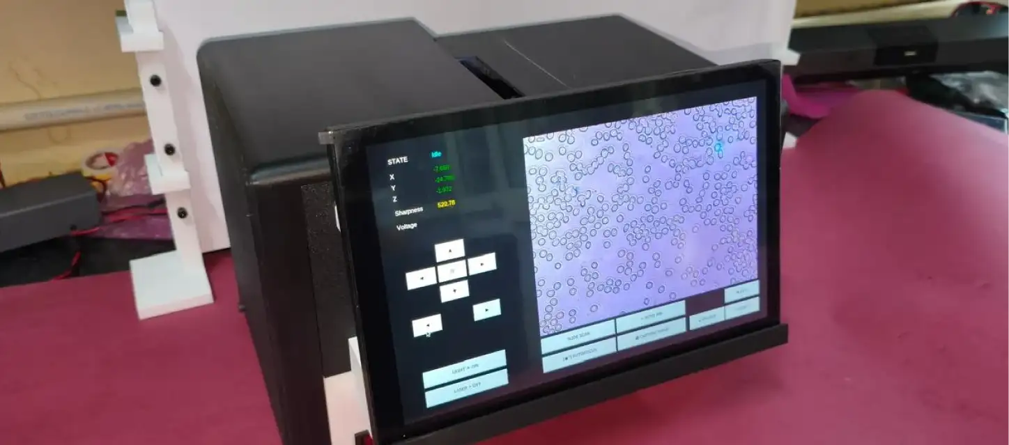
The newly developed optofluidic digital microscopy platform integrates deep learning algorithms with optical imaging and automated motion control systems. The platform continuously analyses microscopic images in real time and automatically adjusts focus through an AI-driven feedback mechanism.
The system was developed at a cost of approximately Rs 1.20 lakh and demonstrated successful laboratory-scale testing in applications including Acute Lymphoblastic Leukemia detection, malaria diagnosis, and complete blood cell count analysis using five-class and seven-class blood cell categorisation models.
The research team included Prof. Earu Banoth, Dr. Shaik Ahmadsaidulu, Amol Lalchand Salve, and Padmanaban Selvakumar.
The platform includes AI-powered autofocus, automated motion control, real-time image processing, cloud-enabled learning capabilities, and imaging support for biological and micro-scale samples. The researchers said the design also focuses on improving repeatability and operational efficiency with minimal human intervention.
Speaking on the project, Prof. Banoth said the team aims to develop a handheld microscopy system capable of delivering performance comparable to imported automated microscopy technologies while supporting multiple biomedical applications.
The researchers are currently working on generating additional ground-truth data and scaling the technology for field deployment and validation across diagnostic centres and research laboratories. The next phase will focus on obtaining feedback for regulatory approvals and commercialisation efforts.
The project received research support from the Anusandhan National Research Foundation, the Department of Science and Technology, and the Department of Biotechnology.
Potential applications of the technology include digital pathology, tissue imaging, AI-assisted microscopy, portable diagnostic systems, laboratory automation, point-of-care diagnostics, and microfluidic analysis.
Stay tuned for more such updates on Digital Health News