New 4D Flow MRI Promises Breakthrough in Diagnosing Aortic Stenosis
Advertisement

The results, published in Open Heart, showed that 4D flow MRI offered more accurate and reliable measurements of blood flow through heart valves than echocardiography.
Researchers from the University of Sheffield and the University of East Anglia (UEA) have developed an advanced MRI technique that could transform the diagnosis of aortic stenosis, a common and life-threatening heart condition.
The research was conducted in collaboration with several institutions, including the Norfolk and Norwich University Hospitals NHS Foundation Trust, Hospital San Juan de Dios (Spain), University of Chieti-Pescara (Italy), University of Leeds, and Leiden University Medical Center (The Netherlands).
The study was funded by the Wellcome Trust.
Faster, More Accurate Diagnosis
Aortic stenosis affects an estimated 300,000 people in the UK and about 5% of people aged 65 and above in the US.
A single-centre study in India reported that isolated aortic stenosis accounts for about 7.3% of valvular heart disease cases, with degenerative calcific disease being the most common cause (65%), followed by bicuspid aortic valve (33.9%).
The disease causes the aortic valve, the heart’s main outflow valve, to stiffen and narrow, limiting blood flow and leading to symptoms such as chest pain, shortness of breath, dizziness, and fatigue.
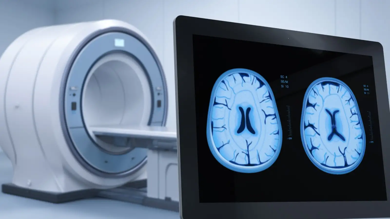
In a recent study, researchers used a novel four-dimensional flow (4D flow) MRI scan to assess 30 patients with diagnosed aortic stenosis and compare its performance against standard ultrasound-based echocardiography. The new technique captures blood flow in three spatial directions over time, allowing for a more comprehensive view of how blood moves through the heart.
The results, published in Open Heart, showed that 4D flow MRI offered more accurate and reliable measurements of blood flow through heart valves than echocardiography.
These findings were further validated by tracking patient outcomes over eight months, revealing that the new method better identified those who needed timely surgical intervention.
Professor Andy Swift, from the University of Sheffield’s School of Medicine and Population Health, said, “4D flow scanning holds significant promise to improve assessment of how patients are affected by aortic stenosis. The enhanced accuracy isn’t just a technical advancement, it may allow for earlier and more precise diagnosis.”
He added that more accurate assessments will empower clinicians to make better-informed treatment decisions and reduce potential complications.
Lead author Dr Pankaj Garg, from UEA’s Norwich Medical School and a consultant cardiologist at Norfolk and Norwich University Hospital, added, “At the moment, doctors use an ultrasound to diagnose the condition, but this can sometimes underestimate the severity of the disease, delaying vital treatment. We wanted to see whether 4D flow MRI could provide a more accurate and reliable diagnosis, and it did.”
With this breakthrough, researchers expect widespread use of 4D flow MRI in clinical practice for earlier and more accurate treatment of aortic stenosis.
Stay tuned for more such updates on Digital Health News.
Stay tuned for more such updates on Digital Health News